Nuclear future under the microscope
Fri, 11 May 2012 13:13:00 BST
New researcher uses the University’s advanced technology to gain vital knowledge of the effects of radiation

As Britain’s nuclear reactors near the end of their lifespan, research at the University of Huddersfield is leading to a greater understanding of the effects of radiation on some of their key components.
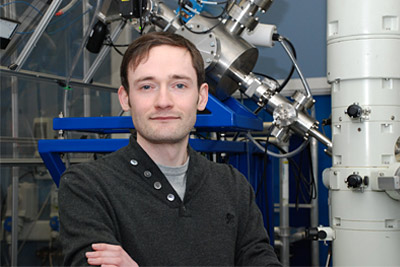
And new research assistant Graeme Greaves (pictured) is able to harness advanced electron-microscopes that enable him to examine materials at the nano-scale. He is one of a team of scientists at Huddersfield and five other universities who have formed the ‘Fundamentals of Current and Future Uses of Nuclear Graphite’ (FUNGraph) Consortium.
“The current generation of nuclear reactors are lasting longer than initially intended,” he says. “They’re approaching the end of their lifetime so we need to understand how the graphite within those reactors is coping with this prolonged use.”
The nuclear industry around the world must know how graphite – used in large quantities in current reactors – is affected by radiation.
Graeme is able to help provide the answer by using lab facilities at the University of Huddersfield that includes an advanced piece of equipment named MIAMI – which stands for Microscope and Ion Accelerators for Materials Investigation.
As a microscope able to examine materials at the nano-scale while bombarding it with an ion beam, it enables researchers to simulate the damage caused to materials by irradiation.
Burnley-born Graeme Greaves – whose doctorate is near completion – comes to Huddersfield from Salford University, the same path taken by his supervisor, Professor Steve Donnelly, the developer of MIAMI, who is now Huddersfield’s Dean of Computing and Engineering.
Graeme is delighted to be taking part in a large-scale research project that has massive real-world implications. And he likes what he has found at Huddersfield.
“The labs are brand new and the facilities are excellent and very well supported. It is a very good research environment.”
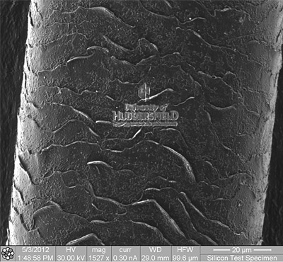
Pictured below is a magnified image of a human hair (~ 60 μm across) that has been engraved with the university's logo, achieved using the University's dual-beam FIB/SEM system.


